Showing posts with label Ortho Supersite. Show all posts

Wednesday, May 5, 2010

Bone Graft substitutes updates

Aesculap Implant Systems launches new demineralized bone matrix 
Aesculap Implant Systems, LLC announced in a press release the recent launch of its ProSpace DBM-D.
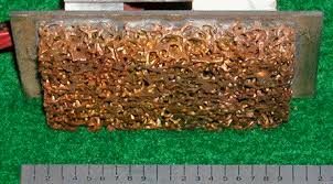
ProSpace DBM-D is a demineralized bone matrix available in two forms, “flowable” paste or “moldable” putty, the company stated The moldable putty contains cortical-cancellous chips which creates a 3-D scaffold for optimized osteoconduction.
ProSpace DBM-D pastes and putties serve as bone void filler in many surgical applications. Unique features associated to this product allow for room temperature storage and re-hydration with a choice of fluids, including patient’s own blood, sterile water or saline.
It is manufactured for Aesculap by RTI Biologics, Inc. 

Using rh-BMP-2 may not guarantee fusion in all cases
Shen HX.Spine. 35(7):747-753. April 2010.
A large consecutive case series of multilevel fusions treated with recombinant human bone morphogenetic-2 yielded a 10.2% pseudarthrosis rate at 6 months.
“Since the risk of pseudarthrosis increases with the number of fusion levels, a long fusion lever arm may biomechanically overwhelm the biologic advantage of rhBMP-2,” the authors wrote in their abstract. “While rhBMP-2 is known to enhance fusion rates, it does not guarantee fusion in all situations.”
Pseudarthrosis rates after anterior cervical fusion range from 0% to 20% for single-level fusions and up to 50% for multilevel fusions, according to the abstract. Some researchers have theorized that rhBMP-2 may decrease the pseudarthrosis rate.
Growing array of bone graft substitutes now available in the United States
Knowing the properties of available bone graft substitutes helps orthopedic surgeons choose appropriate products.
Musculoskeletal allografts are used every day in orthopedic surgery. Last year, more than 1.3 million musculoskeletal allografts were distributed in the United States, according to the American Academy of Orthopaedic Surgeons.
Although use of these biologic materials is common, the field is rapidly changing and orthopedic surgeons need to stay up-to-date on the issues surrounding musculoskeletal allograft tissue.
Among the issues: safety, efficacy, sterilization methods, donor and allograft availability and recall status.
To increase allograft tissue awareness in the orthopedic community, the American Academy of Orthopaedic Surgeons (AAOS) regularly communicates information on the subject to its members via courses and at its Web site (aaos.org).
Earlier this year, the AAOS Orthopaedic Device Forum, Committee on Patient Safety and Committee on Biological Implants Tissue Work Group prepared materials on bone graft substitutes and musculoskeletal allografts for distribution at the AAOS 73rd Annual Meeting.
Among those educational materials was a chart summarizing information about current bone graft substitutes, such as their composition, mechanisms of action and FDA status. With permission from the AAOS, Orthopedics Today is publishing the chart of commercially available bone graft substitutes now available in the United States.
The staff of Orthopaedic Research Laboratories and Lutheran Hospital, a Cleveland Clinic facility, created the chart, contacted all the companies listed and compiled the information about their products.
For more information:
  • Greenwald AS, Boden SD, Goldberg VM, et al. Bone graft subsitutes: facts, fictions and applications. SE#72. Presented at the American Academy of Orthopaedic Surgeons 73rd Annual Meeting. March 22-26, 2006. Chicago.
  • Joyce MJ, Greenwald AS, Boden SD, Heim C, et al. Safety of musculoskeletal allograft tissue. SE #73. Presented at the American Academy of Orthopaedic Surgeons 73rd Annual Meeting. March 22-26, 2006. Chicago.

Despite a variety of bone graft substitute options, an ideal solution eludes surgeons
Understanding available bone graft alternatives and properly selecting them yields optimal results.
by Susan M Rapp
When the acetabulum is at risk for instability or compromised bone growth related to revision arthroplasty, using a bone graft substitute may make good clinical sense, according to a surgeon at the Hospital for Special Surgery.  
Surgeons can choose from materials made of calcium sulfate, calcium phosphate and synthetic substances, as well as a few bone morphogenetic proteins (BMP) and cell-based options, all of which are being used increasingly in hip arthroplasty revision surgery.
However, they vary greatly in terms of their osteoinductivity and osteoconductivity, Bostrom said.

Ultimate substitute

During a presentation at the Current Concepts in Joint Replacement Spring Meeting, Bostrom discussed bone grafting substitute options.
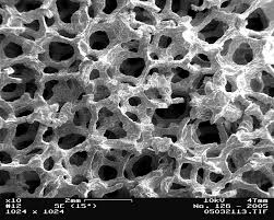
Calcium-based graft substitutes are one option for porous materials, with hydroxyapatite and tricalcium phosphate in forms like pellets, granules and block, among calcium phosphate-based products. These are resorbed over time and new bone forms on top of them.
Plaster of Paris-like calcium sulfate products involve a different biologic process and get dissolved through a chemical process.
Surgeons have also reported success using bone-bank supplied demineralized bone matrix (DBM) in the acetabulum comparable to that of autograft.

Factoring in bone growth

Osteoinductive bone graft substitutes include recombinant human BMP-2 on a resorbable collagen sponge (Infuse, Medtronic Sofamor Danek) and BMP- 7 (Osteogenic Protein-1, Stryker Biotech).
Though mainly indicated for spine and long bone fracture applications, using them in the acetabulum is usually limited to pelvic discontinuities, he said.
Platelet concentration systems that enhance tissue repair factors like fibroblast growth factor and platelet-derived growth fact by four or five times are also available.

For more information:
  • Mathias P.G. Bostrom, MD, can be reached at the Hospital for Special Surgery, 535 East 70th St., New York, NY 10021; 212-606-1000; e-mail: Bostromm@hss.edu. He has no direct financial interest in any product or company mentioned in the article.
Reference:
  • Bostrom MPG. Allograft alternatives: Bone substitutes & beyond. #100. Presented at the 9th Annual Current Concepts in Joint Replacement Spring 2008 Meeting. May 18-21, 2008. Las Vegas.

Monday, April 12, 2010

Treatment of Posttraumatic Deformities in Children and Adolescents by Taylor Spatial Frame

Treatment of Posttraumatic Deformities in Children and Adolescents by Taylor Spatial Frame
By Mark Eidelman, MD; Michael Zaidman, MD; Alexander Katzman, MD
The Taylor Spatial Frame provides outstanding stability and computer accuracy and is a reliable and definite method for posttraumatic deformity correction.
Limb deformity may result from various traumatic sequelae. The most common cause is fracture malunion, but in growing children, shortening and angulation secondary to physis injury is also common. Correction of posttraumatic deformities can be done by various approaches. Each method has pros and cons, but a combination of limb shortening and angulation justify external fixator application.
Several external fixators are available. They can be divided into 2 groups: monolateral and circular. A monolateral external fixator may be more easily tolerated, but it is less stable and forgiving compared to the circular external fixator. The Ilizarov frame is a classic example of a circular external fixator. This frame allows excellent stability but has some disadvantages, such as a long surgeon learning curve and difficulty with rotational correction.The Taylor Spatial Frame (Smith & Nephew, Memphis, Tennessee) is a computerized external fixator with a virtual hinge and the ability to correct 6-axis deformities simultaneously. In contrast with the Ilizarov frame, there is no need for hinge application, multiple frame adjustments, or change of frame configuration to correct multiple plane deformities.
The purpose of this study was to determine the effectiveness of the Taylor Spatial Frame for treatment of complex posttraumatic deformities in children and adolescents.

Materials and Methods

Between 2003 and 2007, 18 patients with various posttraumatic deformities were treated with the Taylor Spatial Frame at our institution (Table). Average patient age at the time of frame application was 13.1 years (range, 8-17 years).
Table: Deformities, Treatment Approaches, and Complications
There were equal numbers of proximal, mid, and distal tibial malunions; 2 patients had combined distal and proximal tibial malunions. Seven patients had deformities secondary to growth arrest: 3 patients had growth arrest of the distal femur, 3 had proximal tibial growth arrest, and 1 had distal radius deformity secondary to physis injury.
Standing anteroposterior (AP) and lateral radiographs from pelvis to toes were obtained pre- and postoperatively and at final follow-up. Deformity analysis and measurements were made in all planes according to the principles described by Paley.1
Surgical technique was described in detail in several reports.1-5 All osteotomies were performed percutaneously by Gigli saw or the drilling and osteotome technique.1 All deformities were analyzed using the total residual correction program and were gradually corrected.
Minimum follow-up was 2 years after frame removal.

Results

In all patients, restoration of the mechanical axis and length equalization was achieved with no or minimal difference compared with anatomical parameters of contralateral extremity. At last follow-up, all patients were pain free and had regained preoperative range of motion (ROM).
The frame was removed after a mean 12.3 weeks (range, 8-24 weeks). Average lengthening was 17.9 mm (range, 5-80 mm).
Eight patients had superficial pin tract infection, which resolved with oral antibiotics or a short course of intravenous administration. One patient had transient peroneal palsy. Another patient had delayed union and needed 2 additional cast immobilizations after fixator removal. The most serious complication was angulation of the regenerate after 40 mm of femoral lengthening (Figure 1). This complication was caused by unstable ring fixation. The fixation block was revised and angulation was successfully and gradually corrected by the total residual program. No patient had deep infection or nonunion.
Figure 1A: A segmented fracture of the femur Figure 1B: Damage of the distal femoral epiphysis Figure 1C: Distal femoral valgus, recurvatum, and external 
rotation Figure 1D: Distal femoral valgus, recurvatum, and external 
rotation
Figure 1E: A segmented fracture of the femur Figure 1F: Damage of the distal femoral epiphysis Figure 1G: Distal femoral valgus, recurvatum, and external 
rotation Figure 1H: Distal femoral valgus, recurvatum, and external 
rotation
Figure 1: Radiographs of a 13-year-old boy with a segmented fracture of the femur and damage of the distal femoral epiphysis (A, B). AP (C) and lateral (D) radiographs before frame application (1 year after trauma) showing distal femoral valgus, recurvatum, and external rotation. AP (E) and lateral (F) radiographs during correction showing regenerate of angulation. Note the distal fragment fixed with 1 ring and only 2 half pins. Radiograph after addition of the second ring and four 1.8 Ilizarov wires (G). Radiograph of normal femoral alignment after frame removal (H).

Discussion

Posttraumatic deformity correction in children with open physis can be a surgical challenge. Proximity of the growth plate restricts the use of intramedullary nail fixation, while shortening and pure soft tissue coverage restrict wide use of the plating technique. The obvious advantage of external fixator application in contrast to internal fixation devices is soft tissue preservation, which can be essential in posttraumatic conditions (Figure 2).
Figure 2A: Severe varus deformity Figure 2B: Pure soft tissue coverage of the medial side Figure 2C: AP radiographs before correction Figure 2D: AP radiographs after correction Figure 2E: Clinical appearance after correction
Figure 2: Photograph of 16-year-old boy with 80-mm shortening and severe varus deformity secondary to damage of the proximal and distal epiphysis (A). Note the pure soft tissue coverage of the medial side of the tibia (B). AP radiographs before (C) and after (D) correction. Clinical appearance after correction (E).
Several external fixators are available. They can be divided into 2 groups: monolateral and circular. A monolateral external fixator may be better tolerated but is less stable and forgiving.6 The stability of the circular frame allows early postoperative weight bearing and ROM maintenance, which can be essential for regenerate formation.2,3 The Ilizarov circular frame is the classic choice for deformity correction and allows correction of almost all possible deformities.7,8 However, correction of multiplanar deformities requires replacement of hinges and frequent frame readjustments. Successful use of the Ilizarov technique has a long learning curve, and correction of complex—especially multiplanar and rotational—deformities remains a difficult challenge, even for surgeons experienced with this technique.3,6
Manner et al9 compared the accuracy of complex deformity correction by Taylor Spatial Frame and Ilizarov circular frame on 208 deformities in 155 patients. They reported that deformity correction was achieved in 90.7% in the Taylor Spatial Frame group vs 55.7% in the Ilizarov frame group. They concluded that the Taylor Spatial Frame has better precision in deformity correction, in 2-, 3-, and 4-dimensional deformity corrections in particular. In most cases, orthopedic surgeons deal with multiplanar posttraumatic deformities.
We treated 18 patients with posttraumatic malunions. Most of our patients had multiplanar deformities and shortening. Despite complex deformities, all patients achieved precise correction of all deformities.
Another choice the surgeon faces is acute vs gradual deformity correction. Matsubara et al10 retrospectively examined clinical results of acute and gradual deformity correction in 2 groups of patients treated by Ilizarov frame or Taylor Spatial Frame. They concluded that gradual correction is a better approach with the use of external fixation.
We believe that gradual correction is a more forgiving and safe way to correct deformities in children. Almost all of our patients had some shortening; therefore, gradual correction with lengthening is the only way to resolve this problem, especially in children with deformities secondary to injury of the growth plate.
We observed relatively few complications in this study. The most common complications were superficial pin tract infections, which were treated with oral antibiotics. There were no deep infections or osteomyelitis. In our previous report,3 the most serious complications were fractures of the regenerate due to pure dynamization in 3 patients. In this study, 1 patient had angulation of the regenerate secondary to unstable fixation of the distal femur. Currently, we use 2 rings at the distal femur with at least one 1.8 Ilizarov wire and three 6-mm half pins.

References

  1. Paley D, ed. Principles of Deformity Correction. Heidelberg, Germany: Springer-Verlag; 2002.
  2. Rozbruch SR, Fragomen AT, Ilizarov S. Correction of tibial deformity with use of the Ilizarov-Taylor spatial frame. J Bone Joint Surg Am. 2006; 88(suppl 4):156-174.
  3. Eidelman M, Bialik V, Katzman A. Correction of deformities in children using the Taylor spatial frame. J Pediatr Orthop B. 2006; 15(6):387-395.
  4. Eidelman M, Katzman A. Treatment of complex tibial fractures in children with the Taylor spatial frame. Orthopedics. 2008; 31(10). pii: orthosupersite.com/view.aspx?rID=31513.
  5. Taylor JC. Correction of general deformity with Taylor spatial frame fixator. J. Charles Taylor Web site. http://www.jcharlestaylor.com/spat/00spat.html. Accessed January 2010.
  6. Binski JC. Taylor spatial frame in acute fracture care. Tech Orthop. 2002; 17(2):173-184.
  7. Ilizarov GA, ed. Transosseous Osteosynthesis: Theoretical and Clinical Aspects of the Regeneration and Growth of Tissue. Berlin, Germany: Springer-Verlag; 1992.
  8. Birch JG, Samchukov ML. Use of the Ilizarov method to correct lower limb deformities in children and adolescents. J Am Acad Orthop Surg. 2004; 12(3):144-154.
  9. Manner HM, Huebl M, Radler C, Ganger R, Petje G, Grill F. Accuracy of complex lower-limb deformity correction with external fixation: a comparison of the Taylor Spatial Frame with the Ilizarov ring fixator. J Child Orthop. 2007; 1(1):55-61.
  10. Matsubara H, Tsuchiya H, Sakurakichi K, Watanabe K, Tomita K. Deformity correction and lengthening of lower legs with an external fixator. Int Orthop. 2006; 30(6):550-554.

Authors

Drs Eidelman, Zaidman, and Katzman are from the Pediatric Orthopedic Unit, Meyer Children’s Hospital, Rambam Medical Center, Haifa, Israel.
Drs Eidelman, Zaidman, and Katzman have no relevant financial relationships to disclose.
Correspondence should be addressed to: Mark Eidelman, MD, Pediatric Orthopedic Unit, Meyer Children’s Hospital, Rambam Medical Center, PO Box 96092, Haifa, 31906 Israel (eidelmanm@gmail.com).
doi: 10.3928/01477447-20100225-16

Excision of Proximal Fibular Tumors: A Newly Described Posterior Surgical Approach

The incidence of primary bone tumors in the fibula is 2.5%.1 The most common tumors found in the proximal fibula are osteochondromas, giant cell tumors, osteosarcomas, and Ewing’s tumors.2
Osteochondromas tend to grow eccentrically rather than centrifugally. Large osteochondromas that continue to grow after skeletal maturity have also been described.3 Osteosarcomas and Ewing’s tumors tend to grow in a centrifugal expansionist fashion, increasing in volume. It is therefore important to note the eccentric location of osteochondromas and their exact relationship to important anatomic structures such as the posterior tibial vessels and nerve, peroneal nerve, anterior tibial vessels, and fibular vessels.
A proximal fibular osteochondroma may distort the normal anatomical course of nerves and vessels and may lead to vascular compression syndromes and a pseudoaneurysm or peroneal nerve paralysis.4 The entrapment of a vessel in the cauliflower-like convolutions of an osteochondroma is also a possibility. A bursa may form about these lesions. An aggressive excision of these proximal tumors may lead to destabilization of the proximal tibiofibular joint.5 Careful staging and planning of the surgical approach and procedure is therefore of the utmost importance in dealing with proximal fibular tumors.
This article describes a surgical approach to deal with posteromedial growth of an osteochondroma that not only allows access and visualization at the posteromedial extension, but also at the anterior extension of such a tumor. At the same time, this approach allows for direct visualization and dissection of the posterior tibial vessels and for an extensive neurolysis of the peroneal nerve.

Case Report

An 18-year-old woman presented with a slowly enlarging posterior left calf mass. She reported exercise- and activity-induced pain with tingling and numbness in the sole of her foot. She had no previous history of tumors, and no one in her family had had any osteochondromas.
Clinical examination revealed a left calf greater in diameter compared to the right calf. The mass was present directly posterior in the calf and was firm in consistency. Both the dorsalis pedis and tibialis posterior pulses were palpable at the ankle. Muscle strength in the posterior tibial and peroneal nerve divisions was normal. No numbness was present during examination.
Radiographs revealed a large mass protruding from the fibula mainly posterior to the tibia (Figure 1). A diagnosis of a large osteochondroma was made. Magnetic resonance imaging (MRI) showed the mass to be extruding from the posteromedial surface of the fibula and extending medially and posteriorly (Figure 2). As this lesion was symptomatic and large, surgical excision was recommended. It was emphasized that nerve and vessel damage were possible. The patient elected to undergo surgery.
Figure 1: Lateral radiograph of an osteochondroma Figure 2: Axial MRI through the proximal fibular tumor
Figure 1: Lateral radiograph of an osteochondroma originating from the posterior aspect of the fibula and extending centrally into the calf muscles. Figure 2: Axial MRI through the proximal fibular tumor, demonstrating the dilemma of using a lateral or medial classic approach to the tumor.
The patient’s recovery was uneventful, with full neurological function without temporary nerve palsy postoperatively.

Surgical Technique

The patient is positioned in the right lateral decubitis position after a spinal anesthetic combined with conscious sedation. A tourniquet is applied on the upper thigh and elevated to 250 mm Hg prior to the incision.
A posterior longitudinal midline incision is used, starting at the flexor crease of the popliteal fossa laterally and extending 14 cm down the calf. Care is taken to preserve both the small saphenous and sural cutaneous nerves. The lateral portion of the incision is developed in the form of a large skin flap deep to the fascia to access the posterior and lateral compartments of the lower leg (Figure 3).
Figure 3: Lateral radiograph of an osteochondroma
Figure 3: Axial transverse anatomical illustration through the proximal third of the tibia with the surgical excision illustrated by dotted lines.
The median raphe of the gastrocnemius muscle is identified and cleaved. The lateral head of the gastrocnemius is carefully dissected loose from the soleus and mobilized laterally. The soleus is detached laterally and retracted medially, hence preserving its innervation on the medial side. The posteromedial part of the tumor can now be dissected free. The fibular attachment of the tumor cannot be accessed from this approach without damaging the lateral gastrocnemius. Therefore, the lateral border of the lateral gastrocnemius is now detached from the posterolateral intermuscular septum, allowing the muscle to be freed and able to be retracted medially or laterally to visualize and access the entire osteochondroma without damage to the lateral gastrocnemius muscle. Care is taken to preserve the proximal vascular supply and innervation of the gastrocnemius. The posterior vessels and nerve are visualized deep to the anterior border of the tumor.
The peroneal nerve is approached with the intent of mobilizing the common peroneal nerve and opening and exposing the common peroneal and deep peroneal nerve branch throughout the fibromuscular tunnel as described by Ryan et al.5 This is necessary to retract the peroneal nerve safely to a more anterolateral position to explore the tumor attachment to the fibula fully. It is imperative to ensure complete mobilization and release of the narrow part of the peroneal nerve through the fibrous tunnel to prevent postoperative compression on the nerve due to reactive swelling.
The next step is to carefully perform a subperiosteal dissection of the anterior periosteum of the tumor’s attachment of the fibula (the tumor stalk).
A curved Homan retractor is placed from superior around the stalk anteriorly to protect the anterior vessels. The tumor stalk is now carefully sectioned with a small oscillating saw. The stalk is retracted posteriorly and its anterior border can now be freed safely by dissection under visualization and protection of the posterior tibial vessels and nerve. Care is taken to remove the entire cartilage cap with its overlying membrane to minimize the possibility of a local recurrence. Sharp spikes of bone protruding from the fibula are smoothed, and visual inspection of the tumor bed as well as of the tumor on the back table is performed. The posterior tibial vessels and the peroneal nerve are inspected to ensure their free passage in the lower leg.
The tourniquet is released and all bleeders secured. The lateral gastrocnemius is sutured back posteriorly to the medial gastrocnemius. A soft drain is placed and the wound closed. A 3-way splint is applied with the ankle plantigrade (neutral) to prevent early muscle contracture and to help with pain management.

Discussion

Malawer2 described 2 types of excisions for tumors of the proximal fibula. The type I excision is wide but more conservative, saving the peroneal nerve and reconstructing the fibular collateral ligament. The type II excision, although also wide, is more aggressive and includes the anterior and lateral compartments, anterior tibial artery, peroneal artery, and proximal tibiofibular joint (en bloc). Both of these excisions are performed through a single incision curvilinear from above the knee, carving anterior to the tibial crest, and ending distal over the peroneal compartment. The flap is based on the posterior (medial) aspect of the skin. This is an excellent approach for centrifugally enlarging aggressive tumors where access to all 3 leg compartments is mandatory. The disadvantage of this incision is the large extent of the dissection to access the posterior compartment and its far medial extension to the medial border of the tibia.
It is for these medially protruding tumors not involving the lateral aspect of the fibula that the described surgical approach was developed. Krieg et al3 reported a case of extensive growth of an osteochondroma in a skeletally mature patient. Axial sections of the MRI showed a posteromedial extension of the tumor up to the medial border of the tibia. It would be difficult to access the entire tumor (similar to our case) from a lateral fibular approach without creating a large skin flap. A posterior midline approach in such cases allows the 2 heads of the gastrocnemius to be retracted sideways, exposing the medial and fibular (lateral) aspect of the tumor safely. If deemed necessary to reach the anterior compartment, it may be accessed by curving the incision anteriorly both at its superior and inferior extents (the reverse of Malawer’s2 skin incision) with its base anterior and lateral.
The incidence of iatrogenic peroneal nerve palsy after removal of fibular tumors is high (4 of 9 cases in the series of Erler et al6 and 3 of 6 type I excision patients of Malawer2). This shows the vulnerability of the common peroneal nerve and its branches after proximal fibular excisions. Palsy may follow excessive retraction or handling of the nerve with metal instruments, incomplete release of the fibular tunnel, and reactive postoperative swelling.
Ryan et al5 performed detailed anatomical dissections of the common peroneal nerve and its branches in the lower leg. They observed the most common site for compression to be the musculoaponeurotic arch at the entrance to the fibular tunnel. In cases of postoperative peroneal palsy, the entrance to the fibular tunnel is typically the area where the nerve is compressed. The deep peroneal nerve may be injured by procedures involving the lateral and anterior aspects of the proximal 8 cm of the fibula. It is therefore imperative to perform a complete release through the fibrous fibular tunnel and to retract the nerve only with soft instruments, eg, a rubber band to prevent iatrogenic peroneal nerve palsy. This is followed by applying adequate soft tissue coverage of the wound and by securing a 30° flexed position of the leg postoperatively.
Popliteal artery entrapment syndrome due to a fibular osteochondroma was described by Guy et al.4 Our patient had similar exertional symptoms due to posterior tibial artery compression. This diagnosis may be easily overlooked, and the claudication symptoms may be ascribed to muscle irritation and other mechanical causes of pain. Careful attention should be paid to preoperative MRI to assess any narrowing or compression of a segment of the posterior tibial vessels.
Tumor volumes >250 mL were reported by Erler et al6 as an indication to sacrifice the deep peroneal nerve to obtain a safe surgical margin. This applies to tumors with a high recurrence rate. Osteochondromas, even >250 mL, may be excised with sparing of the deep peroneal nerve with the caveat that a proper peroneal nerve release is performed.

Conclusion

Excision of benign or malignant tumors of the fibula prove challenging due to the intricacies of the local anatomy with tricompartmental involvement and the proximity of important neuromuscular structures. Careful attention should be paid to the exact anatomical location of the tumor and its involvement of important neurovascular structures in selecting a surgical approach best suited to minimize complications.

References

  1. Unni K. Dahlin’s Bone Tumors: General Aspects and Data on 11,087 Cases. Philadelphia, PA: Lippincot-Raven Publishers; 1996.
  2. Malawer MM. Surgical management of aggressive and malignant tumors of the proximal fibula. Clin Orthop Relat Res. 1984; 186:172-81.
  3. Krieg JC, Buckwalter JA, Peterson KK, el-Khoury GY, Robinson RA. Extensive growth of an osteochondroma in a skeletally mature patient. A case report. J Bone Joint Surg Am. 1995; 77(2):269-273.
  4. Guy NJ, Shetty AA, Gibb PA. Popliteal artery entrapment syndrome: an unusual presentation of a fibular osteochondroma. Knee. 2004; 11(6):497-499.
  5. Ryan W, Mahony N, Delaney M, O’Brien M, Murray P. Relationship of the common peroneal nerve and its branches to the head and neck of the fibula. Clin Anat. 2003; 16(6):501-505.
  6. Erler K, Demiralp B, Ozdemir T, Basbozkurt M. Treatment of proximal fibular tumors with en bloc resection. Knee. 2004; 11(6):489-496.

Authors

Drs Lindeque and Oren are from the Department of Orthopedics, University of Colorado Health Sciences Center, Denver, Colorado.
Drs Lindeque and Oren have no relevant financial relationships to disclose.
Correspondence should be addressed to: Bennie G. Lindeque, MD, PhD, Department of Orthopedics, University of Colorado Health Sciences Center, Mail Stop B202, 4200 E 9th Ave, Denver, CO 80262 (bennie.lindeque@ucdenver.edu).
doi: 10.3928/01477447-20100225-14

Thursday, April 1, 2010

Enzyme may provide quick and accurate diagnosis of periprosthetic joint infections

Posted on the ORTHO SuperSite March 18, 2010
NEW ORLEANS — A strip test indicating the amount of leukocyte esterase enzyme in knee joint synovial fluid following total knee arthroplasty may be a highly sensitive and specific indicator of infected joints, according to the results of a prospective study presented here.
Neutrophils in an infected knee joint secrete the leukocyte esterase enzyme and that the prevalence of this enzyme may be a marker for infection.
Jacovides presented the study at the 2010 Annual Meeting of the American Academy of Orthopaedic Surgeons.
“We believe the leukocyte esterase strip test is a highly accurate test for diagnosis of infection,” Jacovides said. “It is a fast test. It takes 1 to 2 minutes, after which the results are immediately available.”
 They aspirated 1 cc to 2 cc of synovial fluid from 117 TKA cases undergoing revision surgery and applied the fluid to a strip that detected the presence of the leukocyte esterase enzyme. They sent the remainder of the aspirate to be checked for typical counts of leukocyte cells and cultured to determine whether the lab results correlated with the findings of the strip test.
If both tests were positive (++) or if one test was positive (+), the results with the new test were considered positive. All other results were deemed negative, Jacovides said. 
Reference:
Parvizi J, Jacovides CL, Azzam KA, et al. Diagnosis of periprosthetic joint infection: the role of a simple, yet unrecognized, enzyme. Paper #156. Presented at the 2010 Annual Meeting of the American Academy of Orthopaedic Surgeons. March 9-13, 2010. New Orleans.

Staples significantly increase risk of postoperative infection

Posted on the ORTHO SuperSite March 31, 2010
The use of staples to close wounds following orthopedic surgery — especially hip surgery — is associated with a significantly greater risk of wound infection than traditional suturing, according to orthopedic researchers from Norwich, England.
The findings are available at the online home of the British Medical Journal.
Wounds closed with staples were more than three times as likely to develop a superficial wound infection compared to wounds closed with sutures.  In a subgroup analysis of patients undergoing hip surgery, the risk of developing a wound infection was found to be four times greater after staple closure than suture closure, according to the release. 
Staples not recommended
The researchers found no significant difference between staples and sutures in the development of inflammation, discharge, dehiscence, necrosis and allergic reaction.
Reference:
Smith TO, Sexton D, Mann C, et al. Sutures versus staples for skin closure in orthopaedic surgery: meta-analysis. BMJ. [Published online ahead of print March 16, 2010]

Thursday, February 25, 2010

AAOS issues acute Achilles tendon rupture guidelines

The American Academy of Orthopaedic Surgeons recently released a new clinical practice guideline to assist orthopedic surgeons in managing acute Achilles tendon ruptures.
The American Academy of Orthopaedic Surgeons (AAOS) Board of Directors adopted the guideline, The Diagnosis and Treatment of Acute Achilles Tendon Rupture: Guideline and Evidence Report, in December which was endorsed by the American Orthopaedic Foot and Ankle Society.

Acute Achilles Tendon Rupture

File Format: PDF/Adobe Acrobat - Quick View
guideline, The Diagnosis and Treatment of Acute Achilles Tendon Rupture. ... are strongly urged to consult the full guideline and evidence report for this ...
www.aaos.org/Research/guidelines/atrsummary.pdf

Report Title

File Format: PDF/Adobe Acrobat - View as HTML
4 Dec 2009 ... THE DIAGNOSIS AND TREATMENT OF ACUTE. ACHILLES TENDON RUPTURE. GUIDELINE AND EVIDENCE REPORT. Adopted by the American Academy of Orthopaedic Surgeons
www.aaos.org/Research/guidelines/atrguideline.pdf


 Rated recommendations
The recommendations address: what the physical exam for an acute Achilles tendon rupture should entail; the role of imaging in diagnosing these ruptures; the selection of nonoperative and operative treatments; postoperative care; and information concerning return to sports.
In a three-page summary document, the work group that developed the guideline noted that clinicians should read the full guideline and its evidence report. Only then, based on the circumstances presented by the patient, should orthopedists make treatment decisions for these types of injuries.
The AAOS guideline contains 16 recommendations, each of which is rated according to the strength of its recommendation as moderate, weak or inconclusive. Two of the recommendations are based on consensus among work group members, according to the guideline summary appearing on the organization’s Web site.

Postoperative treatment

The most strongly recommended practices, which appear in recommendations numbered 11 and 12, pertain to postoperative treatment protocols and were ranked as moderate strength. In the 11th recommendation, the group wrote, “We suggest early (less than 2 weeks) postoperative protected weight-bearing for patients with acute Achilles tendon rupture who have been treated operatively.”
For the 12th recommendation, they wrote, “We suggest the use of a protective device that allows mobilization by 2 to 4 weeks postoperatively.”
Members of the work group noted in the summary that they were unable to recommend for or against eight practices for acute Achilles tendon ruptures which had inconclusive strength and emphasized that the guideline specifically applies to management of acute Achilles tendon ruptures.

Saturday, February 20, 2010

Shock wave nonunion therapy (ESWT) comparable to surgery in hypertrophic nonunions

Six months after both treatments, radiographic and clinical healing occurred in about 70% of patients. - By Susan M. Rapp
ORTHOPAEDICS TODAY EUROPE 2010; 13:14Extracorporeal shock wave therapy (ESWT)
- provides pain relief
- effective as surgery in resolving hypertrophic nonunions of the femur, tibia and radius.
- noninvasive treatment that focuses pulsed shock waves on targeted areas.
- promote healing by improving the vascularity of tissue and blood supplies.
- "orthopaedic surgeons and their patients now have more options to help repair a fracture that does not respond to initial treatment.”
Extracorporeal shock wave therapy
Healing of various types of nonunion fractures with extracorporeal shock wave therapy was found comparable to surgical results 6 months after both treatments.
Images: Cacchio A
-In study of 126  patients with femoral, tibial or radial nonunions into three groups.
- 1st two groups’ nonunions were treated with ESWT therapy in four sessions, 1 week apart, which each focused 4,000 ESWT impulses on the nonunion site.
-  in group 3 patients received intramedullary nailing, plate fixation or the two combined for their nonunions.
- Based on radiographic and clinical outcomes, the rate of healing within 6 months of treatment was 70%, 71% and 73% in groups 1, 2 and 3, respectively.

- patients examined at 12 and 24 months (after treatment), there were no significant differences in terms of healing. Scans of the bones proved noninvasive shock wave therapy worked just as effectively as surgery”.
Nonunion persistence
Nonunion persisted for 12 months after this 64-year-old woman’s right humerus fracture was fixed surgically.
Fracture callus
A fracture callus is evident in this X-ray of the humerus 6 months after extracorporeal shock wave treatment.
- cases, such as atrophic nonunions or malaligned fractures, where using shock waves may not be as effective.
For more information: Angelo Cacchio, MD, can be reached in the Department of Physical Medicine and Rehabilitation, San Salvatore Hospital of L’Aquila, via L. Natali 1, 67100 L’Aquila, Italy; +39-862-607235; e-mail: angelo.cacchio@tin.it. He has no direct financial interest in any products or companies mentioned in this article.
J. Lawrence Marsh, MD, can be reached at the Department of Orthopaedics, 200 Hawkins Drive, Suite 1181RC, Iowa City, IA 52242-1088 U.S.A. ; e-mail: j-marsh@uiowa.edu. He has no direct financial interest in any products or companies mentioned in this article.
  • Reference:
Cacchio A, Giordano L, Colafarina O, et al. Extracorporeal shock-wave therapy compared with surgery for hypertrophic long-bone nonunions. J Bone Joint Surg (Am). 2009;91:2589-2597.

Metal foam : New material that mimics bone may create better biomedical implants

Orthosupersite
North Carolina State University Researchers developed a “METAL FOAM”  having similar elasticity to bone heading to a new generation of implants that avoid bone rejection.
Characteristics:
- lighter than solid aluminum
- made of 100% steel or a combination of steel and aluminum
- “extraordinarily high-energy absorption capability”
- light weight
- modulus of elasticity similar to that of bone.
- its rough surface  foster bone growth into the implant

Modulus of elasticity

- Modulus of elasticity determines the load bearing of an implant when placed into bone.
- “If the modulus of elasticity of the implant is too much bigger than the bone, the implant will take over the load bearing and the surrounding bone will start to die,”
- “This will cause the loosening of the implant and eventually ends in failure.

Avoiding stress shielding

- modulus of elasticity for the foam is much closer to the 10 to 30 GPa of natural bone than the 100 GPa of titanium.
- light weight of the foam is attributed to its porous nature.
- on ability of the foam to avoid taking on too much load bearing from the surrounding bone “composite foam can be a perfect match as an implant to prevent stress shielding.”
  • References:
Vendra L, Rabiei A. Evaluation of modulus of elasticity of composite metal foams by experimental and numerical techniques. To be published in the March 2010 issue of Materials Science and Engineering A.
 www.ncsu.edu 

Sunday, February 7, 2010

Comparison of Different Lateral Tibia Locking Plates in Schatzker V tibial plateau fractures

A Biomechanical Comparison of Three Different Lateral Tibia Locking Plates
By Bennie Lindeque, MD, PhD; Todd Baldini, MS
ORTHOPEDICS 2010; 33:18

Purpose:  -how well laterally placed modern tibia locking plates used in the treatment of Schatzker V tibial plateau fractures would uphold the medial plateau during axial loading.

- Fifteen third generation Sawbone tibias were obtained and an osteotomy was cut beneath the medial plateau to recreate Schatzker V type plateau fractures. Three groups were created (n=5 per group). Each group was plated with either a Synthes 4.5-mm LCP proximal tibial plate, a Zimmer NCB proximal tibia plate, or a DePuy Polyax tibial plate.
- A vertical load was applied over the medial plateau using an Instron servohydraulic test machine. Load measurements were analyzed at 2 and 3 mm of subsidence as well as load to failure.
- Failure was defined as closure of the wedge osteotomy or the medial condyle collapsing.
- The Synthes and DePuy plates both held up better than the Zimmer plate at 2 and 3 mm of subsidence. Despite this fact, all plates tested held up well above physiological forces of full and partial weight bearing and therefore would be appropriate for the treatment of Schatzker V type tibial plateau fractures

.

The knee is one of the major weight bearing joints of the lower extremity, therefore proper fracture fixation following injury is of the utmost importance.
- These fractures can be divided into 4 groups: distal femur versus proximal tibia and intra-articular versus extra-articular.
-This article focuses on fixation of Schatzker V tibial plateau fractures with large medial defects.1
- Overall, tibial plateau fractures account for 1.2% of all fractures.2
- In the past, this type of plateau fracture has been treated with medial and lateral fixation3-5 or even a combination of internal and external fixation.6-8 But with the advent of locking plates, it has been shown to be as effective in maintaining reduction and stability during healing with unilateral plating as with the traditional methods.9 It has also proven to be an effective technique to offer more fixation strength, which can be of benefit to patients with osteoporotic bone.10
Table 1: Comparison of Lateral Locking Plates
The purpose of this study was to determine whether modern locking plates would be able to uphold a medial plateau fracture above physiological forces.

Lateral Locking Plates
Each company’s plates used different screw configurations, screw sizes, materials, and were of different lengths (Table 1)
Figure 1B: Tibia locking plates Figure 1C: Tibia locking plates
Figure 1: Synthes (A), Zimmer (B), and DePuy (C) lateral tibia locking plates.
Figure 1A: Tibia locking platesThe shortest plate available was used from each company and every hole was filled. If there was a variable angle to the proximal subchondral locking screws, the screws were placed as far apart as possible.

Results

The load data is shown in Table 2. The DePuy Polyax plate had a significantly larger failure load than the Synthes locking compression plates or Zimmer noncontact bridging plates with further analysis. The Synthes locking compression plate and DePuy Polyax plates proved to carry significantly larger loads than the Zimmer noncontact binding plate when looked at with the Tukey-Kramer HSD test.
Table 2: Load Data Mean
The stainless steel Synthes locking compression plates all failed by condyle collapse when the Sawbone fractured at the distal screw. The titanium Zimmer noncontact bridging and titanium DePuy Polyax plates all failed by osteotomy closing without plate breakage.

Discussion

The aim of this study was to ascertain whether 3 different modern locking plates would be strong enough to uphold a Schatzker V fracture under physiological loading conditions and secondarily to directly compare 3 different proximal, lateral tibia plates with locking and nonlocking options in the treatment of Schatzker V type tibial plateau fractures. The use of fully locked and hybrid locking/nonlocking plating systems have been recognized as appropriate treatment of proximal tibial fractures, even with significant bone loss medially.10,12,13

Conclusion

Based on our data, the Synthes and DePuy plates both held up better than the Zimmer plate at 2 and 3 mm of subsidence. Despite this fact, all plates tested held up well above physiological forces of full and partial weight bearing and therefore would be appropriate for the treatment of Schatzker V type tibial plateau fractures.

Read full article:

Thursday, February 4, 2010

Orthopedics Today : Jan-Feb 2010 : Topics of Interest

Orthopedics Today :Feb 2010 : Topics of Interest
1. THA may be beneficial for young patients with Down syndrome and osteoarthritis
Between 8% and 28% of young people with Down syndrome are estimated to have osteoarthritis of the hip. Improved biomaterials and increased life expectancy for this group may make total hip arthroplasty an attractive option for these patients, according to an orthopedic researcher.  

2. Blood transfusions can increase the risk of infection in orthopedic patients
Despite an unknown etiology, transfusion with allogenic blood products predisposes patients to an increased risk of infection. Koval and colleagues reported an infection rate of 27% as opposed to 15% in transfused vs. nontransfused patients undergoing open reduction and internal fixation for hip fracture, out of a cohort of 687 patients. Interestingly, the incidence of urinary tract infection is also considerably higher in patients undergoing orthopedic procedures and receiving blood transfusion, pointing to the possible transfusion-induced immunomodulation (TRIM). 

3. Cytokine biomarkers in orthopedics offer enormous diagnosis and prognosis potentials
finger-width incision the four cytokine biomarkers denote a pathogenic process within a joint correlating to pain in a patient with meniscal pathology. They exist in local inflammatory sites in picomolar concentrations that can increase by thousand-folds in response to pathogenic processes, making them good biomarkers. we found fascinating in our study was the ability of inflammatory cytokines to predict operative pathology. Two patients who had a positive MRI for meniscal pathology were found during arthroscopy to have no significant pathology requiring operative intervention and no evidence of inflammatory cytokines was detected 
4. New minimally invasive technique may be useful to decompress lumbar nerve roots 
Using a new endoscopic technique to treat patients with spondylosis-induced lumbar nerve root compression shows promising results, according to researchers from Japan.“Although spinal fusion is the gold standard to treat spondylolysis and spondylolisthesis, decompression without fusion can be effective procedure for certain patients,”
For the procedure, about a one-finger width skin incision is needed to insert the endoscope.

5. Endoscopy successful in treating snapping iliopsoas tendon

Study in details all articles and more at.....
http://www.orthosupersite.com/
 

New collagen-modifying Osteogenesis Imperfecta gene discovered

1. New collagen-modifying osteogenesis imperfecta gene discovered
Most types of osteogenesis imperfecta (OI), also known as brittle bone disease, that have been identified relate to a dominant mutation in collagen. The type involving the newly discovered Cyclophilin B gene corresponds to a recessive trait, and individuals need two defective copies of the Cyclophilin B gene to develop OI.
The gene is needed to make the protein Cyclophilin B, part of a complex that modifies collagen by folding it into a precise molecular configuration before it is secreted from cells.  
Reference:
Barnes AM, Carter EM, Cabral WA, et al. Lack of Cyclophilin B in osteogenesis imperfecta with normal collagen folding. N Engl J Med. 2010. E-pub ahead of print.
  Read more..

Monday, February 1, 2010

Lumbar Disk Herniation: What Are Reliable Criterions Indicative for Surgery?

Lumbar Disk Herniation: What Are Reliable Criterions Indicative for Surgery?
By Balkan Cakir, MD; Rene Schmidt, MD; Heiko Reichel, MD; Wolfram Käfer, MD
ORTHOPEDICS 2009; 32:589  

Overview

Lumbar disk herniation is the pathologic condition most commonly responsible for radicular pain, and the condition for which lumbar surgery is performed most frequently. This article analyzes the diagnostic findings often considered as reliable criteria for surgical intervention to determine if they are justified by recent literature.